Fig. 2.

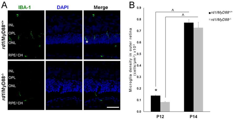
Activated microglia recruitment to the outer retina is reduced at P12 but not at P14 in the rd1/MyD88-/- mouse. (A) Retinal sections from P12 rd1/MyD88+/+ and rd1/MyD88-/- mice immunostained for the activated microglia marker IBA-1 (asterisk), alongside DAPI and merge images. INL= Inner Nuclear Layer, OPL= Outer Plexiform Layer, ONL= Outer Nuclear Layer, RPE/ CH= Retinal Pigment Epithelium/Choroid. Scale bar: 50μm. (B) Microglia cell density in outer retina (from OPL to RPE). Numbers of activated microglia were counted and expressed as number of cells per area (μm2) of outer retina ×103. At P12, there is a 40% reduction in activated microglia in the outer retina of rd1/MyD88-/-compared with rd1/MyD88+/+ mice (n=3 each, p<0.05). Microglia density increases in both mice from P12 to P14, at which point outer retinal microglia numbers are similar in both rd1/MyD88+/+ and rd1/MyD88-/- (n=4, 3 respectively). Mean ± SEM are shown. * p<0.05, comparison among genotypes within a given time-point; (x0005E) p<0.05, comparison between ages of the same genotype.
