
Fig. 3.
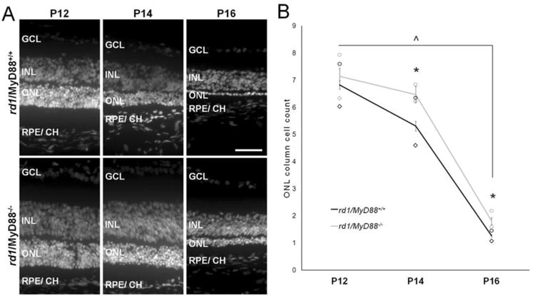
Fewer photoreceptors are lost during retinal degeneration in the rd1/MyD88-/- mouse. (A) Representative IHC sections with nuclear DAPI stain of P12 to P16 rd1/MyD88+/+ and rd1/MyD88-/-retinas revealing increased cells in the outer nuclear layer in the rd1/MyD88-/- across all ages. GCL= Ganglion Cell layer, INL= Inner Nuclear Layer, ONL= Outer Nuclear Layer, RPE/ CH= Retinal Pigment Epithelium/ Choroid. Scale bar: 50μm. (B) Average ONL column counts at P12, P14 and P16 in rd1/MyD88+/+ and rd1/MyD88-/- including averages for central (◊) and peripheral (o) counts. ONL cell number decreases in both genotypes with age, with a 4.9-fold decline from P12 to P16 in the rd1/MyD88+/+ (P12 n=6, P16 n=3, p<0.0001) and a 3.3-fold decline in rd1/MyD88-/- (P12, P16 n=3, p<0.0001). The amount of photoreceptor death was lower at P14 and P16 in the rd1/MyD88-/- mice compared with rd1/MyD88+/+, with 22% more nuclei remaining at P14 (rd1/MyD88+/+ n=4, rd1/MyD88-/-n= 3, p<0.05) and 43% more nuclei at P16 (rd1/MyD88+/+ n=3, rd1/MyD88-/- n= 3, p<0.05) in the rd1/MyD88-/- mice compared to rd1/MyD88+/+. Mean ± SEM are shown. * p<0.05, comparison between genotypes within a given time-point; ^ p<0.05, comparison among ages of the same genotype.