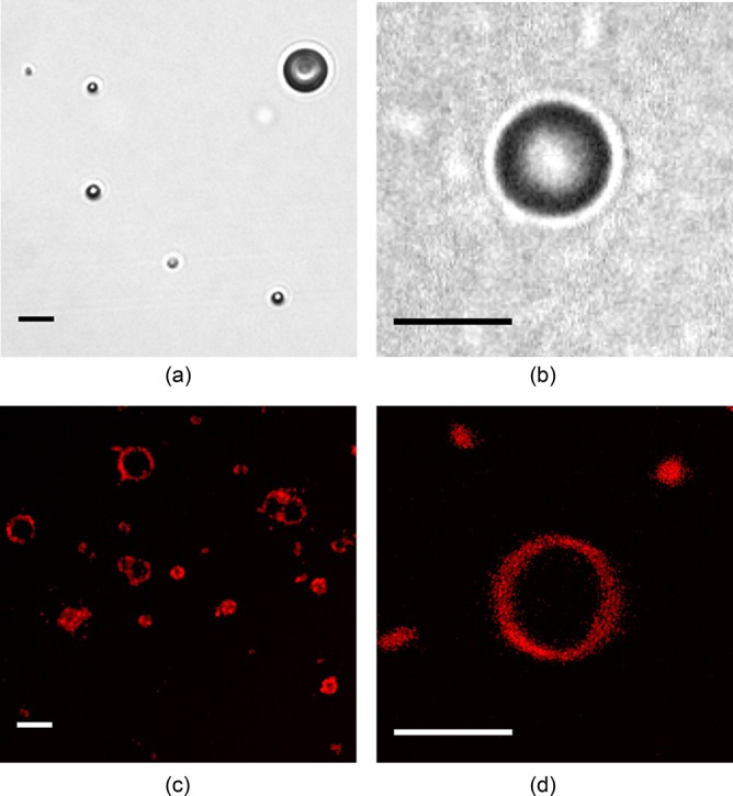
FIG. 2.

(Color online) (a) Optical image of a dilute suspension of ELIP suspended in an OptiCell® (40× magnification). (b) Single frame of a Brandaris 128 recording (120× magnification), (c) and (d) Super-resolution confocal microscope images of fluorescently labeled (2 mol. % rhodamine-DPPE) ELIP in glycerol (Leica TCS 4Pi, 100× magnification). Scale bars represent 5 μm in all images.
