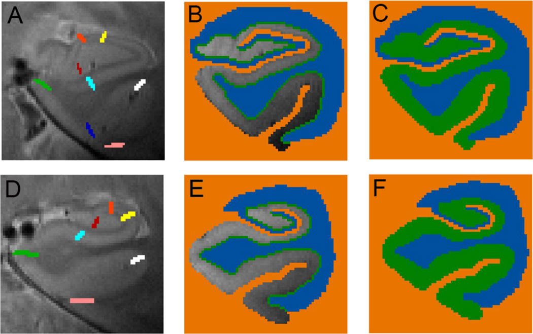
Fig. 4. Unfolding method.
Each subjects’ gray matter (green, C, F) is created by segmenting the lateral and dorsal white matter and CSF areas (blue) and medial non-MTL gray matter and cerebral spinal fluid (orange). The gray matter is grown in layers in between (B, E) from blue to orange voxels. (A, D) Boundaries between regions are demarcated on each slice for each individual subject in 3D space. (A) Shown are the anterior MTL boundaries between dentate gyrus and CA3 (red), CA3 and CA2 (orange), CA2 and CA1 (yellow), CA1 and subiculum (light blue), subiculum and ERC (green), ERC and PRC (dark blue), PRC and fusiform (pink). (D) Additional posterior boundaries are shown including the boundary between subiculum and PHC (green) and between PHC and fusiform (white).

