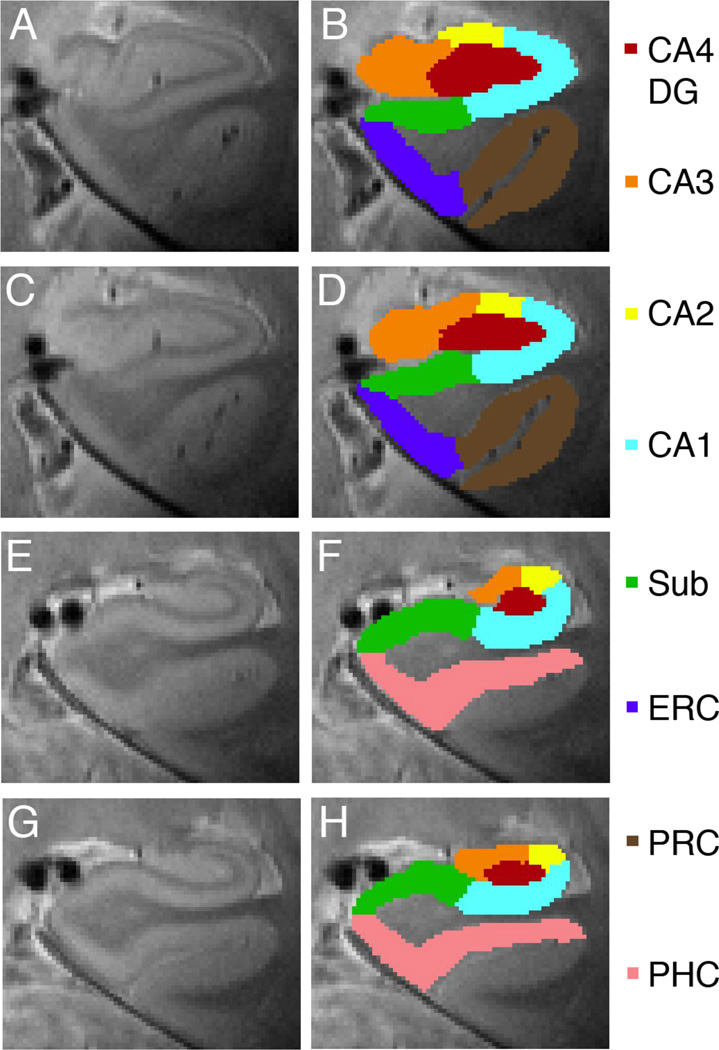
Fig. 5. Anatomical Regions of Interest.
Voxels in 2D space are projected into 3D space to create anatomical regions of interests showing anterior (A–D) and posterior regions (E–H): CA4 and dentate gyrus (CA4DG [red]), CA3 including fimbria (orange), CA2 (yellow), CA1 (light blue), subiculum (green), entorhinal cortex (blue), perirhinal cortex (brown) and parahippocampal cortex (pink).

