
Figure 7. Spatiotemporal regulation of AMPK activity by CID system.
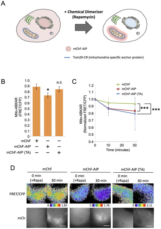
(A) Schematic diagram of CID system. Since cells express anchor protein, Tom20-CFP-FRB (Tom20-CR), diffusive FKBP-fused peptide (mChF-AIP) (left) was trapped at mitochondria in the presence of rapamycin (right).
(B) The effect of AIP and AIP (TA) on AMPK activity at mitochondria. HEK293 cells were transiently transfected with mito-ABKAR, Tom20-CR as anchor protein, and either mChF (n = 39), mChF-AIP (n = 35), or mChF-AIP (TA) (n = 34). Subsequently, AMPK activity at mitochondria was monitored before adding rapamycin. *p < 0.05. N.S., statistically nonsignificant.
(C) Inhibitory effect of sequestered AIP at mitochondria on AMPK activity. AMPK activity at mitochondria in HEK293 cells described in Figure 7B was monitored before (0 minute) and after adding rapamycin at each time points. AMPK activity at each time point was normalized to that in 0 minute. Data are presented as normalized mean ± standard deviation from two independent experiments. ***p < 0.001.
(D) Representative image of Figure 7C were shown. Upper panel: FRET/CFP. Lower panel: mCherry image. Scale bar, 10 μm.