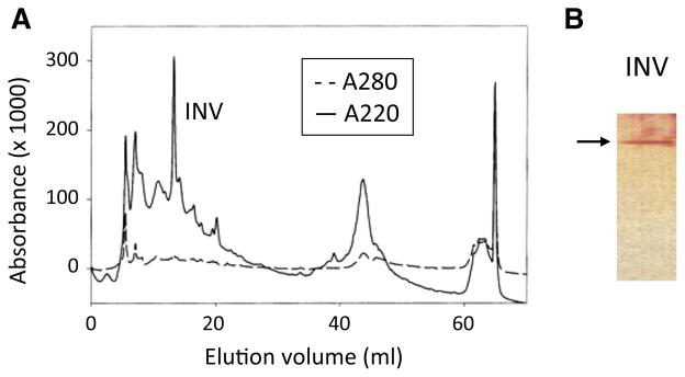
Fig. 2.

Isolation of the L. donovani (1SCL-2D) invertase using FPLC fractionation. Concentrated supernatants from L. donovani (1SCL-2D) promastigote release assays were subjected to FPLC on a Mono-Q column. Individual fractions from this column were collected, optically scanned and their absorbance at 220 and 280 nm were plotted (a). One of these fractions (INV) when subjected to SDS-PAGE and silver staining (b) contained only a single ~70 kDa protein band (arrow). (Color figure online)
