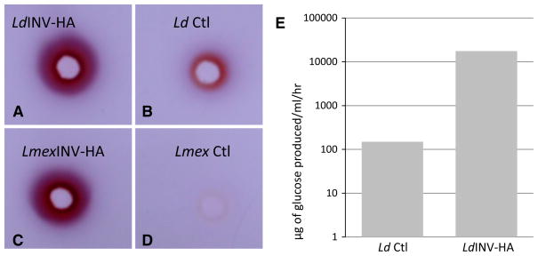
Fig. 8.
Detection of invertase activity produced by INV-HA transfected promastigotes. Aliquots of unconcentrated (1×) culture supernatants from L. donovani LdINV-HA (a) and L. mexicana LmexINV-HA-(c) transfected promastigotes and their respective pKSNEO controls (b Ld Ctl and d Lmex Ctl, respectively) were loaded to precut wells of an agarose gel containing 25 mM sucrose as substrate and allowed to diffuse into the gel for 4 h at 37 °C. Following incubation, the resulting hydrolysis product (fructose) was visualized in these plates using triphenyltetrazolium chloride (TTC) as a capture reagent. The resulting invertase activity is shown in these plates by the presence of a dark red/purple-colored ring of TTC-captured product radiating from the sample wells. The gel shown here is one example of the results obtained from numerous gel assays which were carried out in these experiments. e Quantitation of invertase activity produced by LdINV-HA-transfected promastigotes and their pKSNEO controls. Aliquots of unconcentrated (1×) culture supernatants from LdINV-HA and pKSNEO control promastigotes (Ld Ctl) were assayed using sucrose as substrate in a glucose oxidase spectrophotometric assay. Aliquots of culture supernatants were taken and assayed for invertase activity at a cell density of ~107 parasites/ml. The invertase activity is shown as μg of glucose produced by 107 parasites/ml/hour. (Color figure online)

