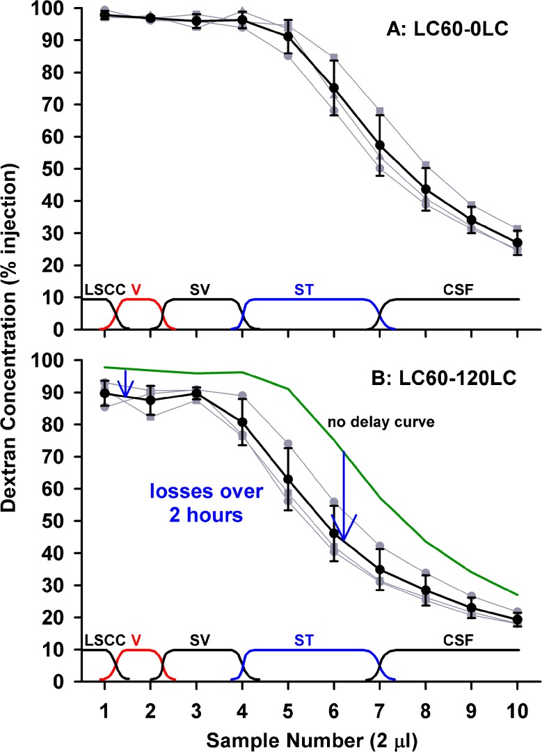
FIG. 1.
Dextran concentrations of 2 μL samples collected sequentially from the lateral SCC either immediately (upper panel) or 120 min after (lower panel) perilymph loading. Individual experiments are shown gray and mean ± standard deviation curves are shown black. Approximate sites of origin of the samples are indicated at the bottom of each plot. With no delay, initial samples were close to the injected concentration, but later samples declined as CSF entry increasingly contributed to the samples. With 120 min delay, initial samples were lower but samples 5–7 were markedly lower. These data are consistent with the greatest decline of dextran occurring at the cochlear apex or in scala tympani.

