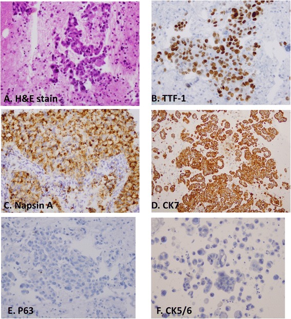
Figure 2.

Immunostaining pattern of TTF-1, Napsin A and CK7 in adenocarcinomas. A, histomorphology of ADC on H&E slide; B, immunostain of TTF-1 in tumor cells, C, immunostain of Napsin A in tumor cells, D, stain of CK7 in tumor cells, E, stain of P63 in tumor cells, and F, stain of CK5/6 in tumor cells. Tumor cells of ADC are positive for TTF-1, Napsin A and CK7, but negative for P63 and CK5/6.
