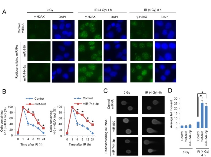
Figure 3.

DSB repair delay by radiation sensitizing miRNAs. (A) Immunofluorescent staining of γ-H2AX foci (green) in DU145 cells transfected with control, miR-890 or miR-744–3p miRNAs in untreated (0 Gy) or 1 and 8 h after IR (4 Gy) treatment. Nuclei were stained with DAPI (blue). (B) Quantification of γ-H2AX foci. The percentage of cells containing >10 γ-H2AX foci (mean ± SE, n = 3) is reported for each time point and treatment group. *, P < 0.05. (C) Comet assay of DU145 cells transfected with control, miR-890 or miR-744–3p miRNAs which were either untreated (0 Gy) or 4 h after IR (4 Gy) treatment. (D) Quantification of the average tail moment (mean ± SE, n = 50) is reported for each miRNA and treatment condition. *, P < 0.05.
