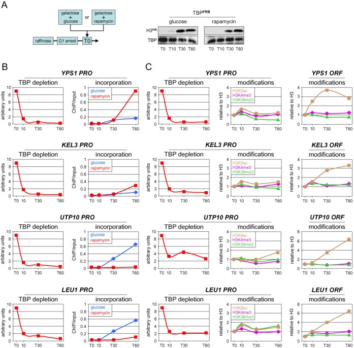
Figure 7.

Loss of TBP from active promoters impacts differently on histone turnover and H3 modifications. (A) Cells from a TBP-anchor-away strain (26) carrying the galactose inducible H3HA construct were arrested in G1 as before. Galactose was then added at T0 to induce expression of H3HA together with rapamycin to deplete TBPFRB from the nucleus (rapamycin). As a control, H3HA expression was induced without TBP depletion by replacing rapamycin with glucose (glucose) to limit galactose induction of the tagged histone when TBP remains nuclear. Shown on the right is a Western blot analysis for H3HA expression under both conditions. (B) TBP promoter occupancy and H3HA incorporation under TBP-depletion (rapamycin, red lines) or control (glucose, blue lines) conditions were monitored by quantitative ChIP using antibodies against TBP and the HA tag. The TBP ChIP values are relative to those measured at T0 for each gene, which were arbitrarily set to 9.0. Results for H3HA incorporation are expressed as the percentage of input DNA recovered. Note that TBP occupancy drops very rapidly, reaching close to background levels already at T10 (min) and that the delay in H3HA incorporation is likely due to the slow kinetics of galactose induction (A). (C) TBP occupancy and the absolute levels of the indicated histone H3 modifications were monitored under TBP depletion conditions using antibodies against TBP and the relevant histone H3 modifications. The ChIP signals (normalized to H3) for H3K4me3 (right panels, violet), H3K9ac (brown) and H3K36me3 (green) are relative to those measured at T0, which were set to 1 to facilitate comparison. (B) and (C) are from two separate experiments. An independent biological experiment is shown in Supplementary Figure S10.
