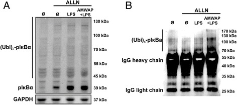
Figure 7.

AMWAP does not inhibit IκBα phosphorylation and ubiquitination. (A) Control and AMWAP-treated BV-2 microglia were preincubated with the proteasome inhibitor ALLN (100 μg/ml) for 30 min to allow for accumulation of phosphorylated IκBα before stimulation with LPS (50 ng/ml) for 30 min. Levels of phosphorylated IκBα were assessed in cytoplasmic extracts using Western blot analysis. AMWAP did not reduce LPS-induced IκBα phosphorylation. The presence of various high molecular weight bands was noticed, presumably representing polyubiquitinated, phosphorylated IκBα. (B) IκBα was immunoprecipitated from cytoplasmic samples of cells treated as in (A). Thereafter, anti-ubiquitin immunoblot revealed the presence of high molecular weight polyubiquitinated forms of phosphorylated IκBα, which were not diminished in AMWAP-positive cells. GAPDH served as loading control. (p)IκBα, (phosphorylated) inhibitor of kappa B alpha; Ubi, ubiquitin; IgG, immunoglobulin G; ALLN; N-acetyl-Leu-Leu-Norleu-al.
