Figure 2.

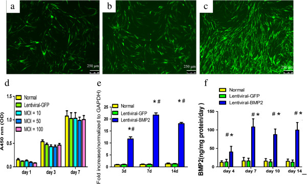
Lentiviral-BMP2 transduced USCs and the effect of transduction on USCs proliferation and BMP2 gene and protein expression. USCs with Lentiviral-BMP2 vectors at different MOIs (a:10; b:50; c:100). Cells were assessed for the presence of positive GFP fluorescence microscopy three days after transduction. Scale bar = 250 μm. (d) Cell proliferation assays were performed at 1, 3 and 7 days post-transduction. The means and standard deviations were calculated from three experiments. (e) RT-PCR analysis showed that the transduced USCs highly expressed the BMP2 gene at days 3, 7 and 14 after transduction. (f) ELISA results indicated that BMP2 production in Lentiviral-BMP2 transduced USCs was significantly increased as compared to that in Lentiviral-GFP transduced USCs or normal USCs. #, P <0.01 (compared with normal USCs) and *, P <0.01 (compared with Lentiviral-GFP transduced USCs). BMP2, bone morphogenic proteins 2; MOI, multiplicity of infection; USCs, urine-derived stem cells.
