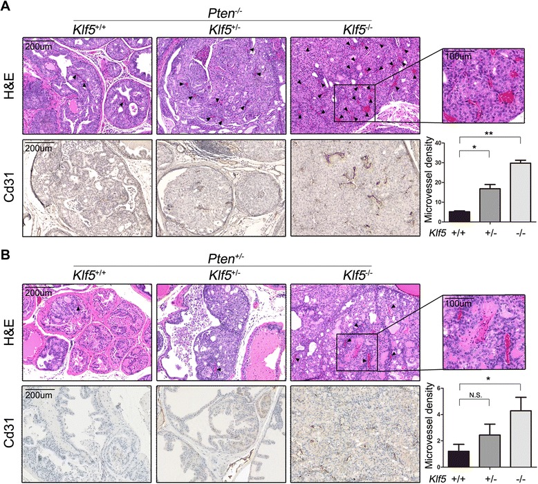
Figure 1.

Klf5 deletion increases blood microvessels in mouse prostate tumors. H&E staining and IHC staining of Cd31 for blood microvessels from prostates at 8 months for Pten −/− (A) and 12 to 18 months for Pten +/− (B). Black arrows point to microvessels inside prostatic acini. Images at the right upper corner are higher magnifications of boxed areas. Microvessels were counted in dorsal lobes for Pten −/− prostates and the entire lobes for Pten +/− prostates. The total area of prostatic epithelia excluding the lumen space inside the acini was measured with ImageJ software. Gene status is indicated at the top of each panel, and + and – indicate wildtype and deletion of Klf5 or Pten, respectively. Three to 6 mice were used for each group. Microvessel density was defined by the number of microvessels per mm2. *, P < 0.05; **, P < 0.01; N.S., not statistically significant.
