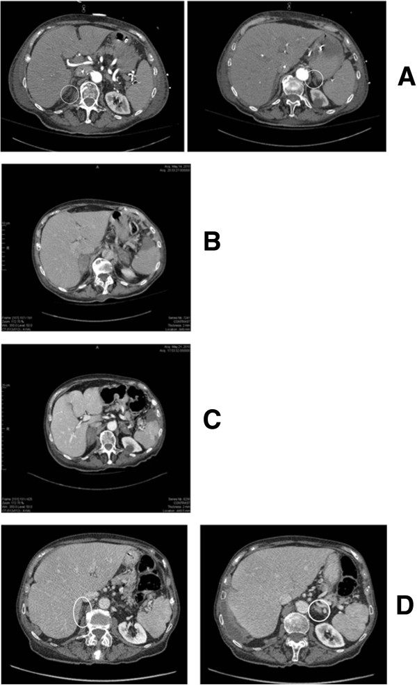
Figure 2.

Abdominal computed tomography scans. (A) Abdominal computed tomography scan on day 3 showing normal adrenal glands. (B) Abdominal computed tomography scan on day 14 showing infiltration with increased adrenal gland volume. (C) Abdominal computed tomography scan on day 21 showing bilateral adrenal hemorrhagic infarction. (D) Abdominal computed tomography scan on day 435 showing small atrophic adrenal glands.
