Figure 2.
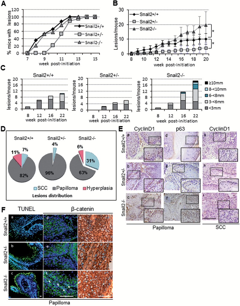
Effect of Snail2 constitutive deletion in DMBA/TPA skin carcinogenesis. (A) Incidence and time of appearance of first lesions in Snail2 +/+ (n = 13), Snail2 +/− (n = 10) and Snail2 −/− (n = 13) mice. (B) Tumor burden indicated as the mean number of lesions/mouse in the three Snail2 genotypes. (C) Tumor size represented as the mean number of lesions per mouse reaching the indicated diameter at week 8, 12, 16 and 22 post-initiation. (D) Distribution in percentage of lesions in Snail2 +/+, Snail2 +/− and Snail2 −/− mice classified as hyperplasia, papilloma or SCC. (E) Immunohistochemical analysis of cyclin D1 and p63 of Snail2 +/+, Snail2 +/− and Snail2 −/− papillomas (a–f) and SCC (g–i). Bar, 500 µm. Amplified images of the indicated areas (squares) are included as insets. (F) Apoptosis (a–c) measured by terminal deoxynucleotidyl transferase-mediated dUTP nick end labeling assay and β-catenin immunofluorescence (d–f) and immunohistochemical (g–i panels) staining of Snail2 +/+, Snail2 +/− and Snail2 −/− papillomas. Nuclei were stained with 4′,6-diamidino-2-phenylindole (blue). White arrows (f) and arrowheads (i) denote cytoplasmic and/or nuclear localization of β-catenin in Snail2−/− papillomas. Bar, 100 µm.

