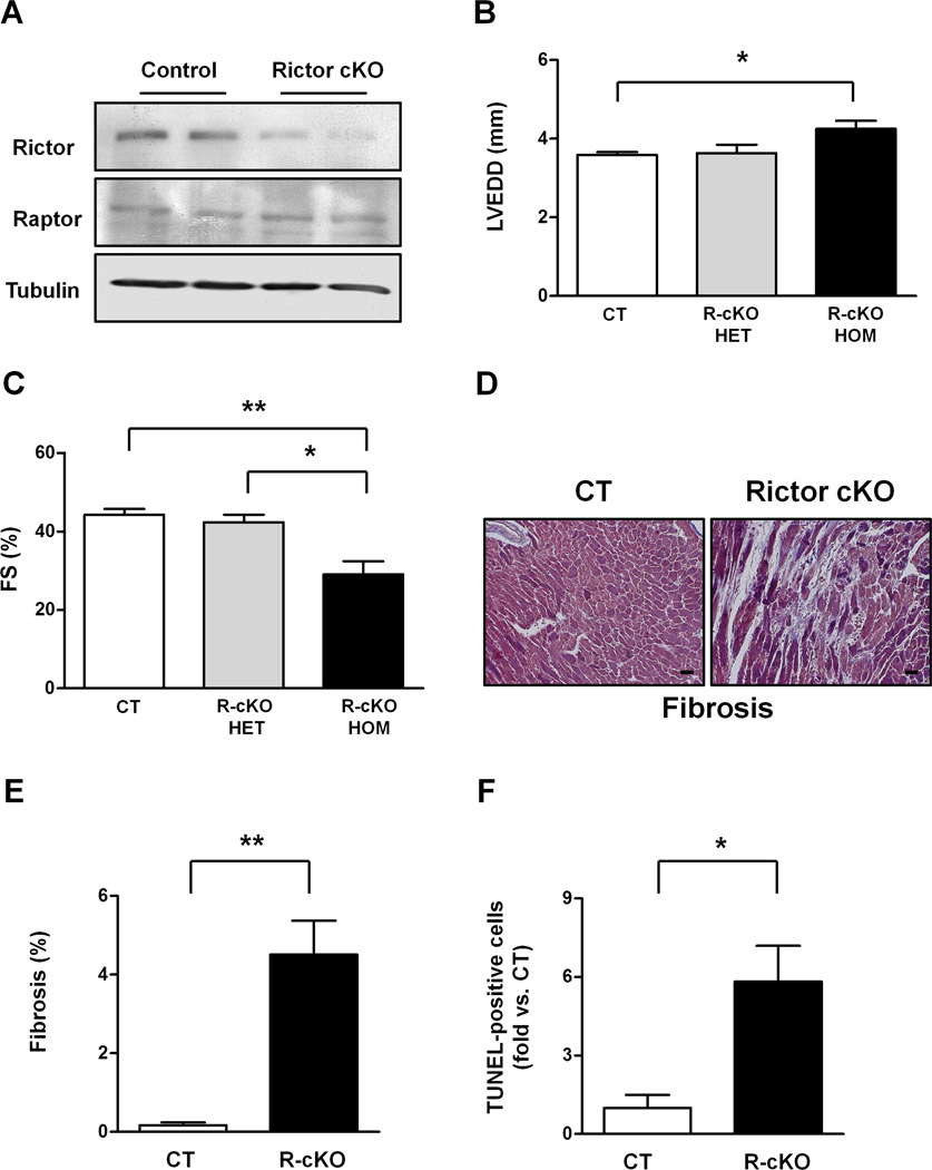
Figure 1. Rictor/mTORC2 disruption promotes progressive cardiac dysfunction and dilation.
A. Representative immunoblot showing Rictor protein levels in control (CT) and Rictor knockout mice (R-cKO). B–C. Six-month-old CT and R-cKO mice (heterozygous and homozygous) underwent echocardiographic analysis. Left ventricular end-diastolic diameter (LVEDD) and fractional shortening (FS) were measured. N=3–8. D–E. Cardiac Masson’s trichrome staining was performed in left ventricular sections from CT and R-cKO mice. Representative pictures (D) and fibrosis quantification (E) are shown. N=4. Scale bar= 50 µm. F. The percentage of TUNEL-positive cells was evaluated in the left ventricle of CT and R-cKO mice. N=3. All data are expressed as mean ± SEM * p<0.05; **p<0.01.

