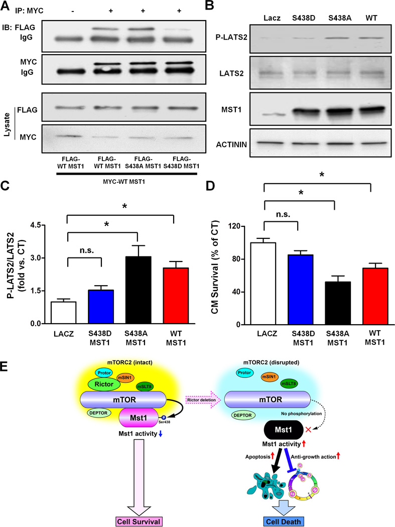
Figure 6. MST1 phosphorylation at serine 438 reduces its dimerization and activity.
A. COS7 cells were transfected with the specified plasmids. MYC was immunoprecipitated and immunoblots for MYC and FLAG are shown. Immunoprecipitate with control IgG was used as a control. B–C. Cardiomyocytes were transduced with ad-LacZ, ad-FLAG-WT-MST1, ad-FLAG-S438A-MST1 or ad-FLAG-S438D-MST1. After 48 hours, the levels of phospho-LATS2 (T1041 homologous), total LATS2 and MST1 were evaluated and representative immunoblots are shown (B) together with quantification analysis of the P-LATS2/LATS2 ratio (C). N=3. D. After 72 hours, cardiomyocyte survival was assessed by CellTiter-Blue assay. N=4. All the data are expressed as mean ± SEM * p<0.05; n.s.= not significant. E. Schema representing the negative regulation of MST1 by mTORC2, which preserves cell survival.

