
FIGURE 5. Rictor−/− DC-secreted pro-inflammatory cytokines, principally IL-6 and IL-23, are key to promotion of Th1/Th17 responses.
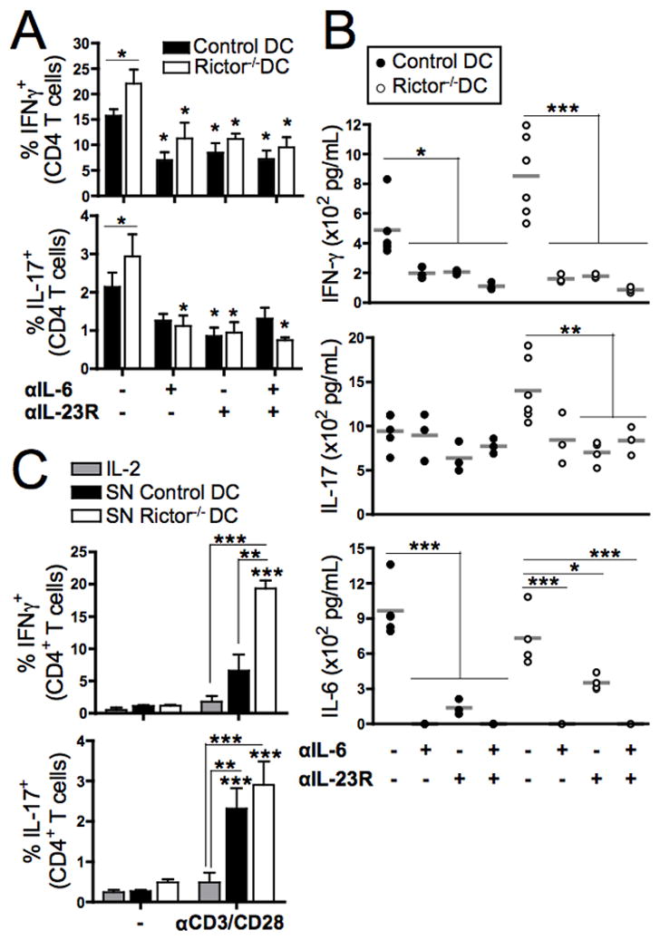
(A) LPS-stimulated control or Rictor−/− DC were used as stimulators of CD3+ BALB/c T cells in 6d MLR, in the presence of blocking mAb against IL-6, IL-23R or both. The CD4+ T cells were then re-stimulated with PMA/Io and stained intracellularly for IFN-γ and IL-17. Bars indicate means + 1SD of n = 3–5 independent experiments, and symbols indicate differences relative to the non-blocked condition. (B) Cytokines in the MLR supernatants were quantified by cytokine bead array on 6d. (C) CD3+ BALB/c T cells were cultured with either IL-2 or DC supernatants, and stimulated with CD3/CD28 beads or left unstimulated (−) for 5d. T cells were re-stimulated with PMA/Io and CD4+ stained intracellularly for IFN-γ or IL-17. Bars are means + 1SD of n = 3–5 independent experiments. Statistical significances are shown compared to the non-stimulated condition. * P < 0.05, ** P < 0.01, *** P < 0.001.