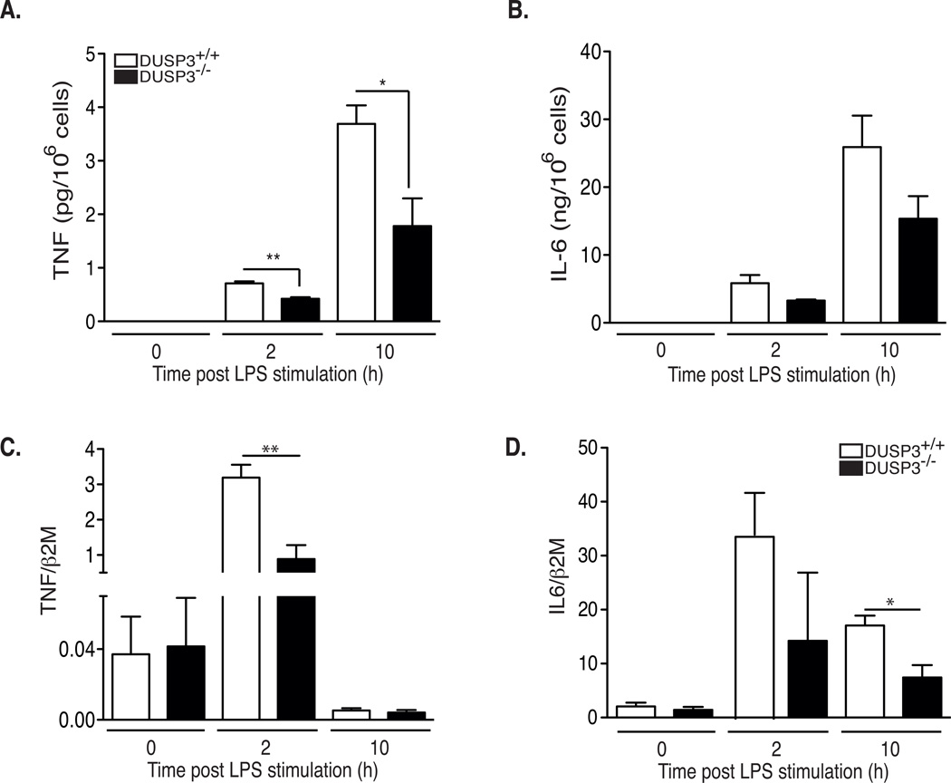
Figure 4. DUSP3−/− macrophages produce less TNF than DUSP3+/+ in response to LPS.
(A–B) DUSP3+/+ and DUSP3−/− peritoneal macrophages were activated ex vivo with LPS (1µg/mL). Cells supernatants were collected at 0h, 2h and 10h after activation. TNF (A) and IL6 (B) concentration were determined using CBA. (C–D) IL-6 and TNF relative to β2M mRNA expression levels in peritoneal macrophages of DUSP3+/+ and DUSP3−/− activated ex vivo with LPS (1µg/mL) for 2h and 10h. Results are presented as mean ± SEM. *P<0.05, **P<0.01. Three macrophage pools prepared from three mice each were analyzed separately.

