Figure 1.
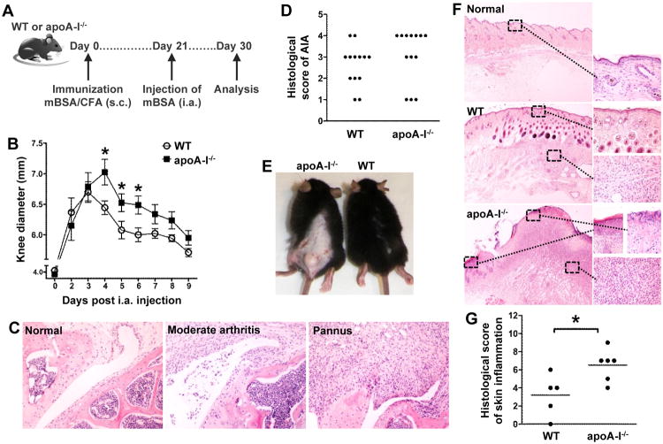
ApoA-I-/-mice demonstrate an aggravated AIA phenotype. (A) WT and apoA-I-/-mice (n=13/group) were immunized subcutaneously (s.c.) with mBSA/CFA and arthritis was induced upon intra-articular (i.a.) injection of mBSA 21 days later. (B) Evaluation of arthritis by measurement of knee joint swelling daily after i.a. injection (n=8-9/group) (day 4: *p=0.0233, day 5: *p=0.0455, day 6: *p=0.0182). Results are expressed as mean ± SEM. (C) Representative H&E stained sections of inflamed joints from WT (moderate arthritis) and apoA-I-/- mice (pannus) as well as non-injected knee joint (normal) are shown (original magnification ×200). (D) Histological scores for AIA in WT and apoA-I-/- mice. The histopathologies of injected knee joints were scored as described in Methods;*p=0.0393 for pannusvs non-pannus.(E) Photos of skin lesions in apoA-I-/- mice 21 days after mBSA immunization. (F) Representative H&E stained sections of control and inflamed skin (original magnification: left side ×20, right side ×400). (G) Histological grading of skin inflammation developed in WT and apoA-I-/- mice (*p=0.0239). In all panels, the data are from two independent experiments.

