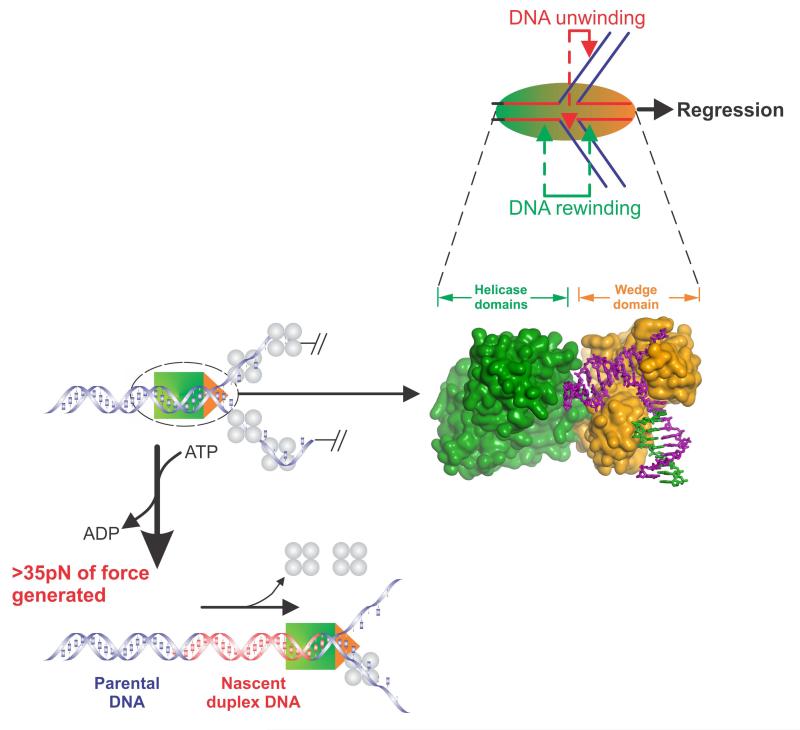
Figure 5. Components of the fork regression reaction catalyzed by RecG.
Top panel, RecG (sphere) is shown bound to a fork in the process of catalyzing regression in the direction of the black arrow. During this reaction, the enzyme couples the hydrolysis of ATP to the simultaneous DNA transactions of unwinding of the nascent heteroduplex regions (red arrows) and DNA rewinding that occurs ahead of the translocating enzyme as well as in its wake (green arrows). Bottom left panels, RecG is shown in the process of fork regression on a DNA substrate bound by the single stranded DNA binding protein. During regression, RecG generates greater than 35pN of force that is sufficient to displace the bound SSB protein. Middle panel, Connolly surface of RecG bound to a model fork. The helicase and wedge domains are coloured to match the schematics in the other panels. When viewed in this way, it is clear that the wedge domain is bound at the fork while the helicase domains translocate along duplex DNA.

