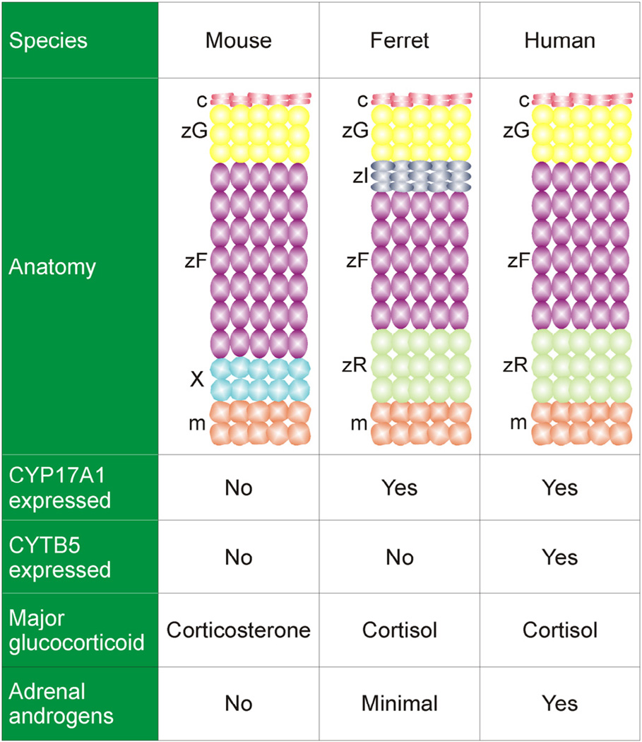
Fig. 1.
Comparative anatomy and physiology of the adrenal cortex. The mouse adrenal cortex contains a zG and zF, but there is no recognizable zR. The adrenal cortex of the young mouse contains a transient layer, the X-zone. The ferret adrenal cortex has an additional layer, the zona intermedia; the analogous layer in the rat houses stem/progenitor cells (Guasti et al., 2013b). CYP17A1 is a bifunctional enzyme with Q both the 17α-hydroxylase activity required for the synthesis of cortisol and the 17,20-lyase activity required for the synthesis of androgens. Cyp17a1 is expressed in the fetal but not postnatal adrenal cortex of the mouse, so under normal conditions the mouse adrenal secretes corticosterone as its major glucocorticoid and does not produce androgens. CYTB5 selectively enhances the 17,20-lyase activity of CYP17A1 through allosteric effects. Non-neoplastic adrenocortical cells in the ferret lack CYTB5, which may account for the low production of adrenal androgens in healthy ferrets. Abbreviations: c, capsule; m, medulla; X, X-zone; zF, zona fasciculata; zI, zona intermedia; zG, zona glomerulosa; zR, zona reticularis.

