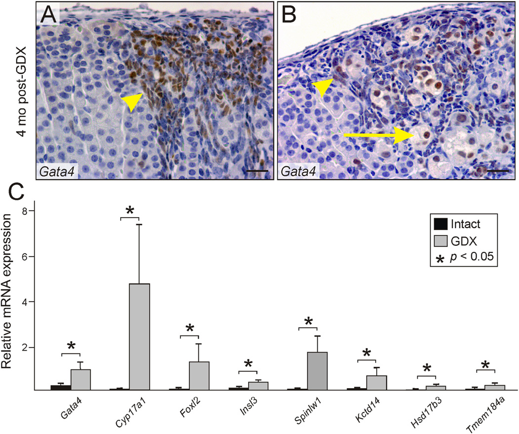
Fig. 2.
Changes in histology and gene expression associated with GDX-induced adrenocortical neoplasia. (A,B) Neoplastic cells in the adrenal cortex of a B6D2F1 female mouse that underwent prepubertal GDX 4 mo earlier. Immunoperoxidase staining for GATA4 highlights small type A cells (arrowheads) and large type B cells (arrows). Bars = 100 µm. (C) Expression of gonadal-like differentiation markers in the adrenal glands of gonadectomized vs. intact female DBA/2J mice. Whole adrenal glands from 4-mo-old gonadectomized or intact virgin female DBA/2J mice were subjected to qRT-PCR analysis. Results are normalized to β-actin mRNA levels (× 102). *P< 0.05.

