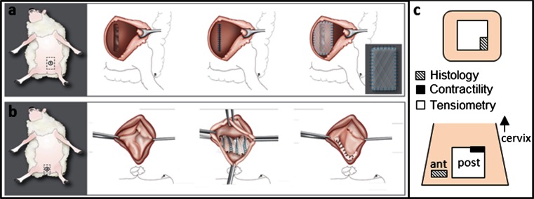
Fig. 1.
Schematic drawing of abdominal (a) and vaginal (b) implantation in the sheep model. Specimens explanted (c) from the abdomen and anterior (ant) and posterior (post) vaginal wall were divided according with their respective testing method. The arrow is pointing cranially in the direction to the uterine cervix (illustration by Myrthe Boymans)

