Fig. 1.
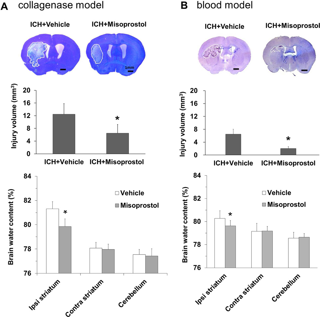
Misoprostol treatment decreases brain lesion volume, edema, and neurologic deficits on day 3 after ICH. Representative images of Luxol fast blue/Cresyl Violet-stained brain sections on day 3 after ICH in the collagenase model (A, top) and in the blood model (B, top). The lesion area, circled in white, is indicated by a lack of staining. Brain lesion volume, which was corrected for brain swelling, was smaller in the misoprostol-treated group than in the vehicle-treated group in the collagenase model (n = 11 mice/group, A, middle) and in the blood model (n = 8 mice/group, B, middle). Brain water content was less in striatum of misoprostol-treated mice than in striatum of vehicle-treated mice in the collagenase model (n=5 mice/group, A, bottom) and in the blood model (n = 5 mice/group, B, bottom). Scale bars: 1 mm. All values are means ± SD; *p<0.05, **p<0.01, t-test.

