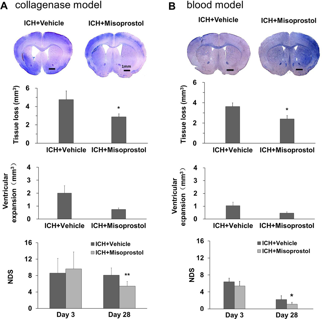
Fig. 2.
Misoprostol treatment decreases brain tissue loss, ventricular expansion, and neurologic deficits on day 28 after ICH. Representative images of Luxol fast blue/Cresyl Violet-stained brain sections on day 28 after ICH in the collagenase model (A, top) and in the blood model (B, top). Residual lesion, circled in white, can be clearly observed in the collagenase model. Compared with vehicle treatment, misoprostol treatment decreased volume of brain tissue loss and ventricular expansion in both models. Misoprostol also ameliorated neurologic deficit score (NDS) on day 28, but not on day 3, after ICH in the collagenase model (n = 8 mice/group, A) and blood model (n = 6 mice/group, B). Scale bars: 1 mm. All values are means ± SD; *p<0.05, **p<0.01, t-test.

