
Figure 5.
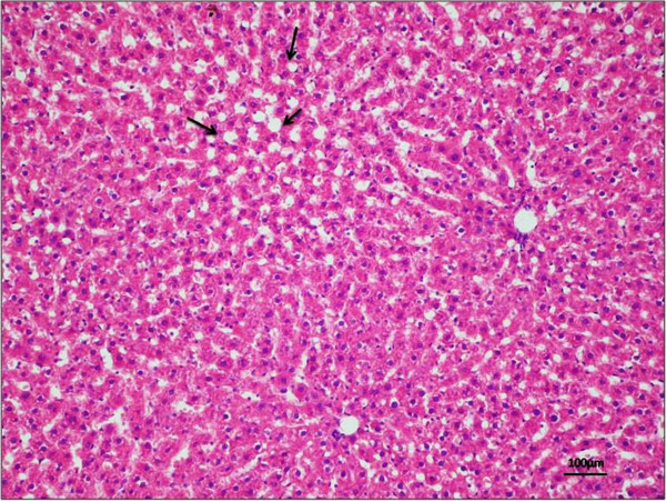
Morphological and histological analysis of a liver sample. H&E staining in liver tissue demonstrated no sign of tissue degeneration, with a good preservation and the beginning of formation of lipid droplets (black arrows). NAFLD activity score = 1 for steatosis, 0 for lobular inflammation, 1 for hepatocellular ballooning and 0 for fibrosis. Magnification × 20; scale bar: 100 μm.