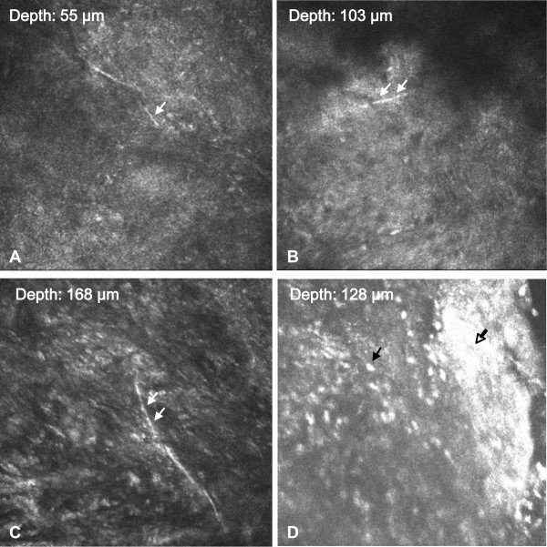
Figure 2.

In vivo confocal microscopy examination. (A~C) Images from different depth show hyper-reflective branching hyphae-like bodies (white arrow) could be identified in the cornea. (D) Infiltration of inflammatory cells (black arrow) and necrotic tissues (hollow arrow). Magnification: ×800.
