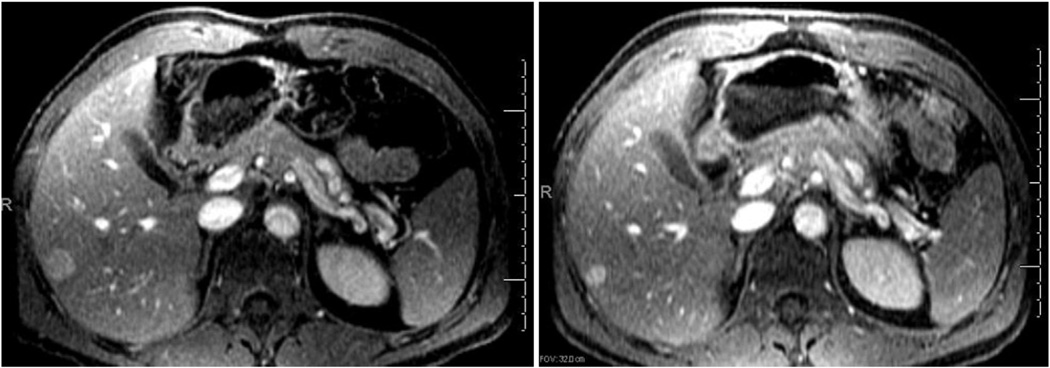
Figure 2.
Selected MR images from a patient with numerous hepatic metastases involving more than 20% of the liver at baseline (left) and 6 months after immunoembolization (right). The patient showed a PR in multiple hepatic metastases, and the largest lesion in the right hepatic lobe decreased from 1.9 × 1.8 cm to 1.2 × 1.0 cm. No progression of hepatic metastasis was seen for 13.8 months.

