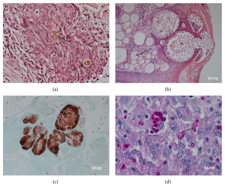
Figure 1.
Photographs of representative fungi-containing samples analyzed in this study. (a) Case 1. Chromoblastomycosis of the skin. Fungal elements can be recognized in HE-stained sections as brownish-yellow pigmented bodies (arrows). A separation of the thick-walled fungus is also visible (multiform body). (b) Case 9. Skin lesion in rhinosporidiosis. Numerous spherical structures varying in diameter in an age-dependent manner. Immature forms (trophocytes) contain nucleus (arrow) and cytoplasm. Mature forms (sporangia) contain numerous endospores (asterisk). The rupture of this form is also visible. HE staining. (c) Case 10. Mycetoma. Lobulated grain with light colored center (brown) in lung. Grocott stain. (d) Case 11. Histoplasma infection. The periodic acid-Schiff reaction reveals many ellipsoidal yeast cells (red) inside macrophages and a giant cell. The arrow shows a budding.

