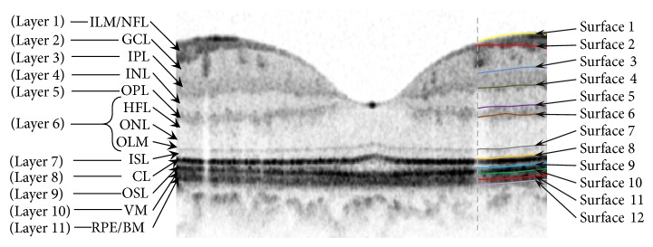
Figure 2.

Segmentation on the original image. Retinal layers are as follows: inner limiting membrane (ILM), nerve fiber layer (NFL), ganglion cell layer (GCL), inner plexiform layer (IPL), inner nuclear layer (INL), outer plexiform layer (OPL), Henle's layer (HFL), outer nuclear layer (ONL), outer limiting membrane (OLM), inner segment layer (ISL), connecting cilia (CL), outer segment layer (OSL), Verhoeff membrane (VM), retinal pigment epithelium (RPE), and Bruch membrane (BM).
