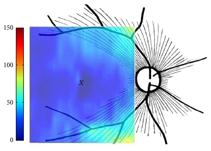
Figure 9.

Agreement between schematic representation of retinal radial peripapillary capillaries in man (X = fovea) (reproduced from [50]) and the calculated RNFL thickness. The fibers in the temporal part of the retina (corresponding to the nasal visual field) course away from the fovea and then once in the nasal retina the fibers turn back toward the optic disc, entering in the superior and inferior portions of the disc. The fibers crowded with higher thickness in superior and inferior areas as they become closer to optic disk.
