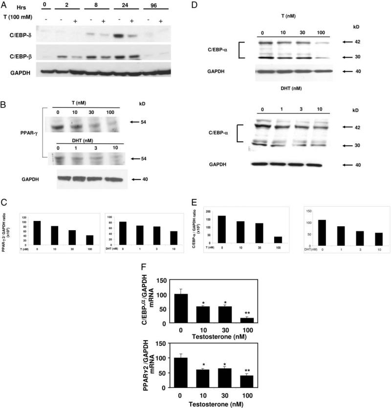
Fig. 3.
Time course of C/EBP-β and C/EBP-δ expression during adipogenic differentiation with and without androgens and dose-dependent inhibition of C/EBP-α and PPAR-γ expression in 3T3-L1 preadipocytes by androgens. A, Confluent 3T3-L1 cells were treated with 100 nm testosterone (T) and allowed to differentiate in AM for various time points. Cells were harvested, lysed, and 100 μg of total protein was electrophoresed on SDS-containing polyacrylamide gels, and Western blot analysis was performed using anti-C/EBP-β, anti-C/EBP-δ, or anti-GAPDH antibodies. Three independent experiments were conducted and data from one representative experiment are shown. B, Top panel, 3T3-L1 cells were treated with either testosterone (0–100 nm) or DHT (0–10 nm) for 12 d and allowed to differentiate in AM as described in Materials and Methods. Cells were harvested, lysed, and 100 μg of total protein was electrophoresed on SDS-containing polyacrylamide gels, and Western blot analysis was performed using anti-C/EBP-α, anti-PPAR-γ, or anti-GAPDH antibodies. Three independent experiments were conducted and data from one representative experiment are shown. C, Densitometric analysis of the PPAR-γ expression (B, top panel) in 3T3-L1 cells after various doses of androgens normalization with GAPDH. D, 3T3-L1 cells were treated with testosterone (0 –100 nm) or DHT (0 –10 nm) and allowed to differentiate in AM as in panel B for 12 d. Cells were harvested, lysed, and 100 μg of total protein was electrophoresed on SDS-containing polyacrylamide gels, and Western blot analysis was performed using anti-C/EBP-α or anti-GAPDH antibodies. Three independent experiments were conducted and data from one representative experiment are shown. E, Densitometric analysis of the expression of C/EBP-α protein (panel C) in 3T3-L1 cells with various doses of androgens after normalization with GAPDH. F, Total cellular RNA from 3T3-L1 cells was prepared using Trizol reagent after incubation with graded concentrations of testosterone (0–100 nm) for 12 d. Quantitative analysis of C/EBP-α and PPAR-γ2 mRNA levels was performed by real-time RT-PCR after normalizing with GAPDH using gene-specific primers. (Significance: *, P < 0.02; and **, P < 0.005 compared with the control group.)

