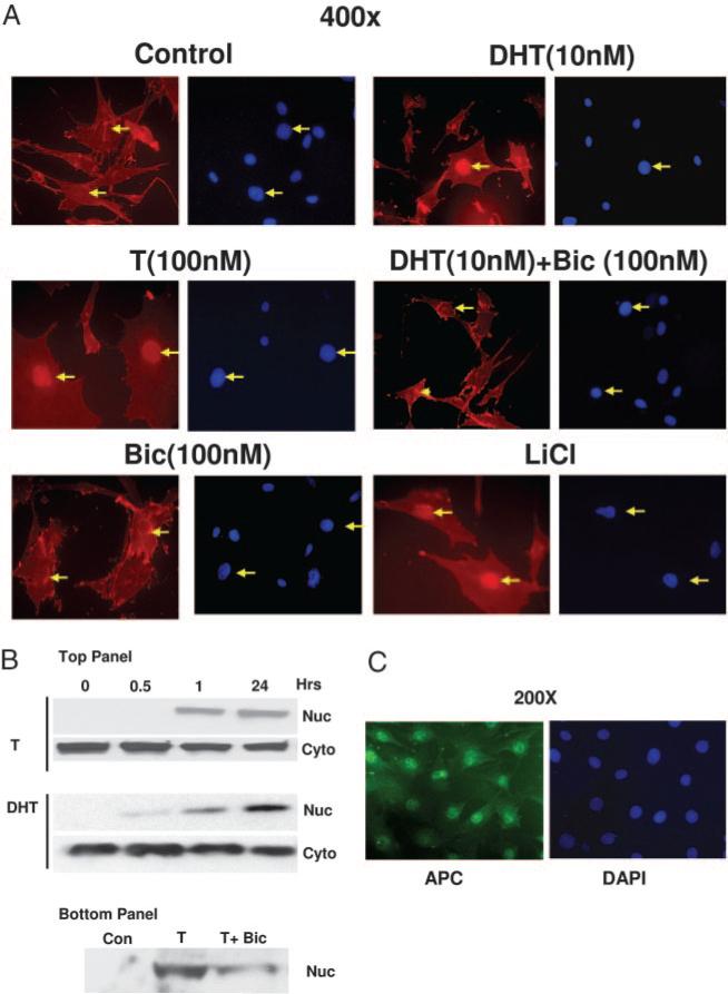
Fig. 4.
AR mediated nuclear translocation of β-catenin in 3T3-L1 preadipocyte cells after testosterone or DHT treatment. A, Cells were grown for 24 h on eight-well chamber slides at 40% confluence and fixed for 20 min with 2% paraformaldehyde after various treatments, as shown. Localization of β-catenin was detected by using anti-β-catenin antibody and Texas Red-conjugated secondary antibody (red). The cells were counter stained with a nuclear stain DAPI (blue). B, Top panel, Cells were treated with T (100 nm) or DHT (10 nm) for various time points (0–24 h) and nuclear and cytoplasmic fractions were analyzed by Western blot analysis using anti-β catenin antibody. Bottom panel, Cells grown with none (control), T (100 nm) or T+ Bic (300 nm bicalutamide) for 24 h, nuclear and cytoplasmic fractions were analyzed by Western blot analysis using anti-β catenin antibody. C, Cells were treated as described in panel A, and localization of APC was detected by using anti-APC antibody and FITC conjugated secondary antibody. The data for androgen treatment groups are not shown. The arrows show the location of the nuclei. The cells were also counterstained with DAPI (blue).

