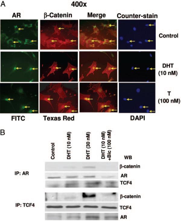
Fig. 5.
Colocalization and physical interaction of AR and TCF4 with β-catenin in DHT-treated cells. A, Double immunofluorescence: Cells were treated as described in Fig. 4 and double immunofluorescence experiments were performed using anti-AR and FITC-labeled secondary antibody for the detection of AR (green) and Texas Red-conjugated secondary antibody for the detection of β-catenin (red), and merging of the red and green pictures are shown as yellow. Cells were counterstained with DAPI (blue) to localize the position of nuclei in these cells. B, Immunoprecipitation: Cells were treated with either DHT (10 and 30 nm) alone or with bicalutamide (100 nm) for 24 h and immunoprecipitation was carried out using 500 μg of total cell lysates and 1–2 μg of respective primary antibodies (see Materials and Methods). The immunoprecipitated proteins were analyzed by Western blot analysis. Upper panel shows the detection of β-catenin, AR, and TCF4 bands after immunoprecipitation with anti-AR antibody, and lower panel shows the detection of β-catenin and TCF4 bands after immunoprecipitation with anti-TCF4 antibody.

