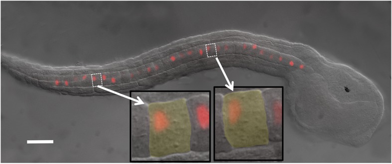
Figure 1. Ciona intestinalis late-tailbud embryo (stage 23) expressing an electroporated Histone 2A/Red Fluorescent Protein (H2A-RFP) in the notochord.
Insets show two cells to illustrate the polarization of the nuclei to the posterior of the cells. Scale bar is 50 μm.

