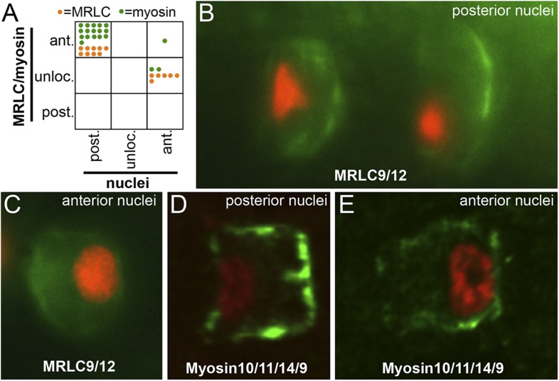
Figure 8. Myosin10/11/14/9 and MRLC9/12 localization in homozygous aim embryos.
(A) Distribution of nuclear and Myosin10/11/14/9 (green) or MRLC9/12 (orange) localization phenotypes (ant., anterior; post., posterior; unloc., unlocalized) in aimless embryos. Each dot represents a single scored cell. (B and C) MRLC9/12-Venus (green) localization in cells with posterior and anterior nuclei (red), respectively. (D and E) Myosin10/11/14/9-myc (green) localization in cells with posterior and anterior nuclei (red), respectively. Anterior is to the right in all panels.

