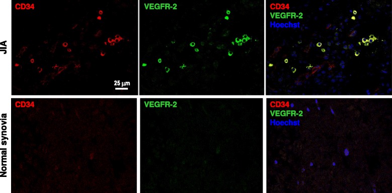
Figure 4.

Bona fide EPC labelling in synovial tissue of JIA patients. A representative confocal imaging analysis of synovial tissue from a JIA patient (upper lane) and from a child who underwent surgical debridement for a non-inflammatory condition (lower lane). The staining for CD34 and VEGFR-2 (KDR) shows double positive CD34+KDR+ cells (bona fide EPCs) in clusters or as isolated cells only in the JIA case, while the control was completely negative. Note that, as expected, CD34 also labels microvessels, although at lower intensity compared to isolated EPCs.
