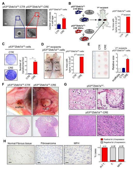
Figure 2.
Lrf loss in MSCs leads to formation of mesenchymal tumors. (A) Soft-agar assay for detecting anchorage independent cell growth of p53KOZbtb7aF/F-CTR and p53KOZbtb7aF/F-CRE MSCs. Representative pictures of the colonies are shown on the left, while the quantification on the right is shown as average of 3 biological independent replicates ± SEM. (B) Schematic overview of the experimental design is shown on the left, while the percentage of mice with a tumor bigger that 0.5 cm3 is shown on the right. p53KOZbtb7aF/F-CTR cells were not transformed, and only scaffolds within mesenchymal cells were recovered after transplantation. (n=2 CTR, n=2 CRE). (C) Detection of transformation status of p53KOZbtb7aF/F-CTR or p53KOZbtb7aF/F-CRE cells. Pictures of the foci are shown on the left, while the quantification of the transformed foci is shown on the right. The quantification on the right is shown as pooled from 3 independent experiments, mean ± SEM. (D) Tumors generated by p53KOZbtb7aF/F-CTR or p53KOZbtb7aF/F-CRE cells transplanted within scaffolds into second recipient mice. Representative pictures of mice are shown on the left, while the percentage of mice with a tumor bigger that 0.5 cm3 (scaffold size used as control) is shown on the right. (n=4 CTR, n=7 CRE) (E) Sizes of tumors generated by p53KOZbtb7aF/F-CTR or p53KOZbtb7aF/F-CRE cells transplanted within scaffolds into second recipient mice. Pictures of the collected tumors are shown on the left, while the relative size of tumors is shown on the right (scaffold size used as control). (F–G) Tumors generated by p53KOZbtb7aF/F-CRE cells compared p53KOZbtb7aF/F-CTR cells collected from 1st recipients, seeded into scaffolds and the transplanted into 2nd recipients. H&E staining showing the morphology of collected tumors or mesenchymal cells on top of the scaffold. p53KOZbtb7aF/F-CTR cells were not able to generate tumors in vivo and only scaffolds within mesenchymal cells were recovered after transplantation, while p53KOZbtb7aF/F-CRE cells originated undifferentiated sarcomas. (* indicates the scaffold. Scale bars: 20 μm). (H) LRF expression in human undifferentiated sarcomas. (M.F.H. malignant fibrous hystocytoma; F. fibrosarcomas; N.F.T. normal fibrous tissue; scale bars 30μm).

