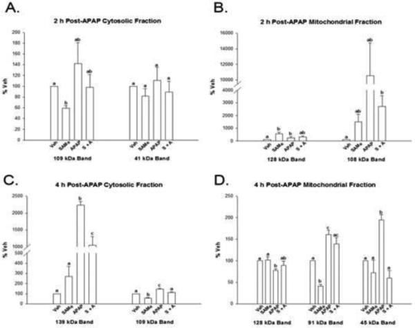
Figure 5. Densitometry analysis of liver mitochondrial and cytosolic 4-HNE adducted proteins 2 and 4 h following APAP treatment.
Densitometry results for Western blots depicted in Figure 4. Panel A (cytosol) and Panel B (mitochondria) depicts results 2 h following APAP overdose. Panel C and D are the cytosolic (C) and mitochondrial (D) fractions examined 4 h following APAP overdose. Groups with a different superscript letter (a, b or c) are statistically different (p<0.05). Cytosolic bands (Panel C) at 139 and 109 kDa bands were highest (p<0.05) in the APAP group when compared to Veh, SAMe and APAP groups. Mitochondrial 4 h samples (Panel D) had the highest intensity in the 45 kDa band compared to Veh, SAMe and S+A.

