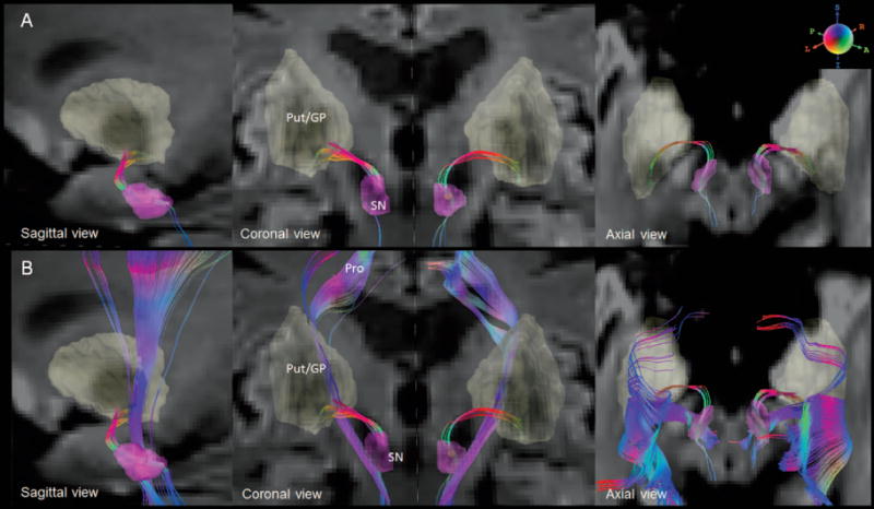
Figure 1.

A. The reconstructed nigrostriatal fibers with color-encoded orientations: green for anterior-poster, red for transverse, and blue for superior-inferior directions. The connecting nuclei are highlighted on diffusion weighted images. B. The nigrostriatal fibers are distinguished from the neighboring fibers (Pro).
SN = substantia nigra (SN), Put/GP = putamen/globus pallidus, Pro = projection fibers
