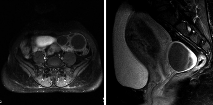
Figure 5.
A 29-year-old female with an invasive mole. the T1 post-contrast fat-saturated axial and sagittal pelvic images show multiple cysts arising from the left ovary in the left iliac fossa without any obvious solid component, and a right ovarian cystic lesion in the pouch of Douglas. There is a heterogeneously-enhancing lesion within the endometrial cavity with invasion of the anterior myometrium and the overlying serosa.

