Figure 2.

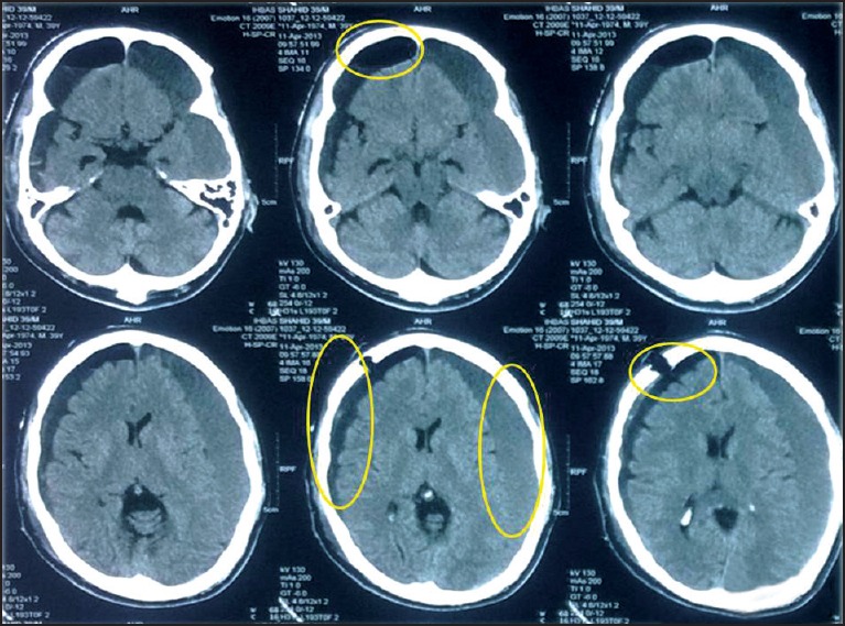
CT scan of head (post-operative) showing the burr-hole, postoperative collection of exudate in the subdural space, pneumocephalus changes, subdural hematoma on the opposite side (nonoperated side-left side), and diffuse cerebral atrophy

CT scan of head (post-operative) showing the burr-hole, postoperative collection of exudate in the subdural space, pneumocephalus changes, subdural hematoma on the opposite side (nonoperated side-left side), and diffuse cerebral atrophy