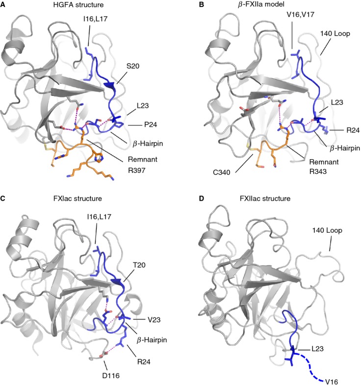
Fig 5.

Cartoon diagrams showing stabilizing interactions in the region of the heavy chain remnant (orange) and the activation loop (blue) are compared for different proteases with salt bridges or hydrogen bonds illustrated as a dotted purple line. (A) Hepatocyte growth factor activator (HGFA) (Protein Data Bank [PDB]: 1YC0), (B) β-FXIIa, homology model, (C) FXIa (PDB: 1XX9), which has no remnant, and (D) the FXIIac structure reported here. The FXIIac activation loop does not have a formed β-hairpin, and residues after Leu23 are not present in the electron density (dotted blue line).
