
Figure 3.
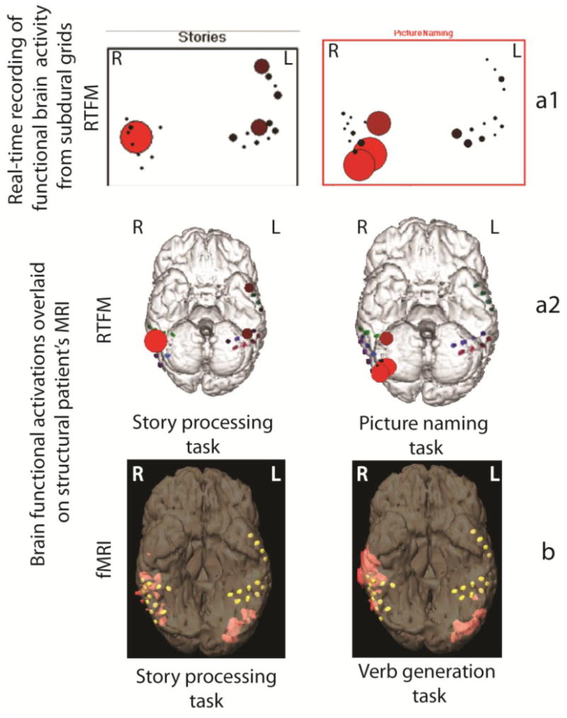
Cortical language activation in epilepsy patient: (a) – results of RTFM recording performed after 1st grid placement procedure. Cortical RTFM activation maps indicate right-sided language laterlization elicited during picture naming and story processing task. The RTFM responses are maximal in the right lateral and basal temporal regions (locations with significant levels of activation are presented as large red circles; grid placement locations are indicated as red, green and blue dots); (a1) – indicates the response recorded from the grids in real-time; (a2) – indicates the same response overlaid off-line on the 3-D model of the patient's brain; (b) – fMRI activation maps with verb generation and story processing tasks (locations with significant levels of activation are indicated in orange; grid placement locations are indicated as yellow dots). With “L” is indicated left hemisphere, with “R” is indicated right hemisphere.