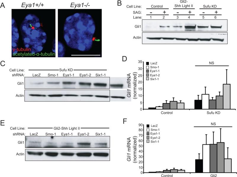
Figure 3.
Eya1 and Six1 function in Shh transduction between Smo and Sufu. (A) Eya1−/− granule cell precursors are ciliated. γ-tubulin (red) marks basal bodies and acetylated-α-tubulin (green) marks the ciliary axoneme; nuclei are visualized by DAPI (blue); scale bar = 10 μm. (B) Stable cell lines with Sufu knock-down (Sufu KD) or Gli2 overexpression (Gli2-ShhLightII) have constitutively elevated levels of Gli1 protein (see lanes 1, 3, 5), Actin = loading control. (C) Eya1 and Six1 shRNAs do not reduce Gli1 protein levels in Sufu KD cells (Actin = loading control). (D) Eya1 and Six1 shRNAs do not reduce Gli1 mRNA in Sufu KD cells (N=5; N=3 Six1-1). (E) Eya1 and Six1 shRNA do not alter Gli1 protein in Gli2-ShhLightII cells (Actin = loading control). (F) Eya1 or Six1 shRNA do not reduce Gli1 mRNA in Gli2-ShhLightII cells (N=5, N=2 Six1-1, Error bars = SEM; NS = not significant, P>0.05).

