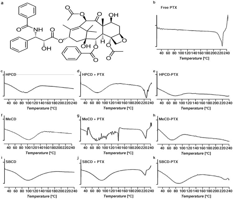
Figure 1.

Representative differential scanning calorimetry (DSC) thermograms of free paclitaxel (PTX), β-cyclodextrins (CDs) and their mixtures and inclusion complexes with PTX; (a) – Chemical structure of PTX. (b) – Free PTX; (c) – Hydroxy propyl β-cyclodextrin (HPCD); (d) Physical mixture of HPCD and PTX (e) – Hydroxy propyl β-cyclodextrin-paclitaxel (HPCD-PTX); (f) – Methyl β-cyclodextrin (MeCD); (g) Physical mixture of MeCD and PTX; (h) – Methyl β-cyclodextrin-paclitaxel (MeCD-PTX); (i) – Sulfobutyl ether β-cyclodextrin (SBCD); (j) – Physical mixture of SBCD and PTX; (k) – Sulfobutyl ether β-cyclodextrin-paclitaxel (SBCD-PTX). A vertical bar on ordinate corresponds to 1 W/g of heat flow.
