Figure 4.

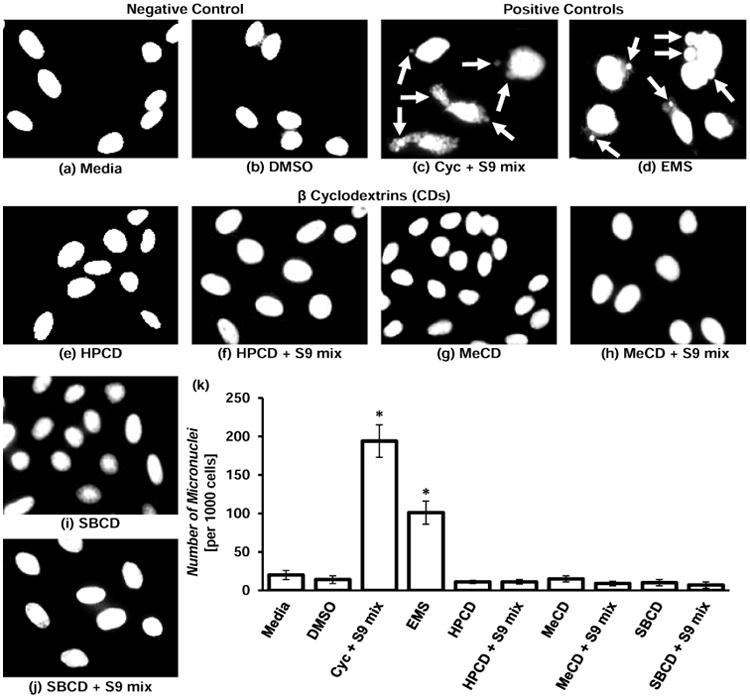
Genotoxicity of β-cyclodextrins (CDs). (a) – (j) – representative images of cells stained with nuclear dye incubated with the following substances: (a) – Media (untreated cells, negative control); (b) – DMSO (negative control for the solvent); (c) – Cyclophosphamide (Cyc) with metabolic activator – S9 mix (positive control 1); (d) – Ethyl methanesulfone – EMS (positive control 2); (e) – Hydroxy propyl β-cyclodextrin (HPCD); (f) – HPCD + S9; (g) – Methyl β-cyclodextrin (MeCD); (h) – MeCD + S9; (i) – Sulfobutyl ether β-cyclodextrin (SBCD); (j) – SBCD + S9. (k) – Quantitative analysis of micronuclei formation. Means ± SD are shown. *P < 0.05 when compared with control (untreated cells). Arrows indicate micronuclei.
